国际生殖健康/计划生育 ›› 2021, Vol. 40 ›› Issue (3): 193-195.doi: 10.12280/gjszjk.20200543

• 病例报告 • 上一篇    下一篇

胎儿脑灰质异位合并FLNA基因突变病例报告

申永梅, 陈叙, 张蕾, 于红, 赵晓敏, 常颖()   

  1. 300100 天津市中心妇产科医院天津市人类发育与生殖调控重点实验室(申永梅,陈叙,常颖),产前诊断中心(张蕾,赵晓敏,常颖),放射科(于红)
  • 收稿日期:2020-09-18 出版日期:2021-05-15 发布日期:2021-05-28
  • 通讯作者: 常颖 E-mail:changying4470@sina.com
  • 基金资助:
    天津市中心妇产科医院/天津市人类发育与生殖调控重点实验室开放基金(2019XH19);天津市自然科学基金青年项目(20JCQNJC00840)

Fetal Heterotopic Gray Matter with FLNA Gene Mutation: A Case Report

SHEN Yong-mei, CHEN Xu, ZHANG Lei, YU Hong, ZHAO Xiao-min, CHANG Ying()   

  1. Tianjin Key Laboratory of Human Development and Reproductive Regulation (SHEN Yong-mei, CHEN Xu, CHANG Ying), Prenatal Diagnosis Center (ZHANG Lei, ZHAO Xiao-min, CHANG Ying), Radiology Department (YU Hong), Tianjin Central Hospital of Gynecology Obstetrics, Tianjin 300100, China
  • Received:2020-09-18 Published:2021-05-15 Online:2021-05-28
  • Contact: CHANG Ying E-mail:changying4470@sina.com

摘要:

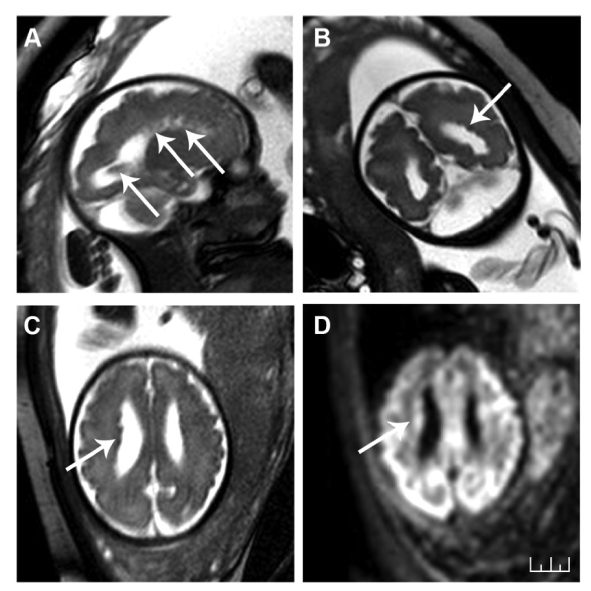
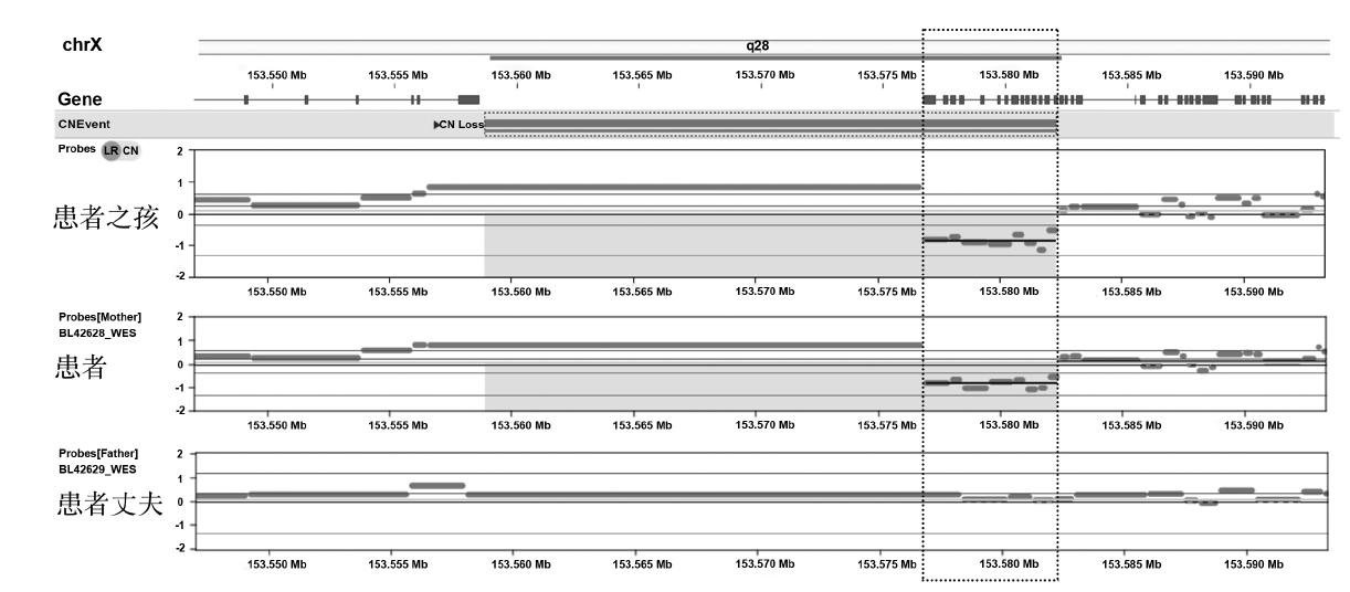
报告1例产前超声联合胎儿颅脑磁共振成像(magnetic resonance imaging,MRI)诊断胎儿脑灰质异位合并细丝蛋白A(filamin A,FLNA)基因突变的病例。患者超声检测结果显示侧脑室外壁凹凸不平,颅后窝池宽,透明隔间腔略窄,小脑延髓池轻度增宽。MRI诊断结果为胎儿脑灰质异位合并大枕大池。羊水穿刺获得脱落细胞,采用全外显子测序和基因拷贝数变异(copy number variations,CNV)分析,显示与脑灰质异位疾病相关的FLNA基因外显子出现缺失,其为可能导致脑灰质异位的致病基因。因此,超声结合胎儿颅脑MRI能有效提高脑灰质异位的诊断率,其中染色体检查可筛查出部分致病原因,并对下次妊娠提供帮助。该病例可丰富临床医生对胎儿大脑灰质异位的产前超声检查、合并畸形、临床表现等的认识。

关键词: 脑室旁小结异位, 室管膜, 超声检查,产前, 磁共振成像, 产前诊断, 病例报告

Abstract:

This paper reported a case of fetal heterotopic gray matter with FLNA gene mutation. Fetal heterotopic gray matter was diagnosed by the prenatal B ultrasound combined with fetal brain MRI. The ultrasound examination showed that the external wall of lateral ventricle was uneven, the posterior fossa cistern was wide, the diaphragmatic space was slightly narrow, and the cerebellomedullary cistern was slightly widened. The gray matter heterotopias and large occipital cistern were affirmed by MRI. The extoliated cells in amniotic fluid was harvested, and whole exon sequencing and CNV analysis were used to test those pathogenic genes of heterotopic gray matter disease. The results showed the deletion of FLNA gene exon. Therefore, the ultrasound combined with fetal brain MRI could effectively improve the diagnosis rate of heterotopic gray matter, and chromosome examination could screen out some pathogenic causes and provide help for the next pregnancy. This case can enrich our understanding of prenatal ultrasound examination, the complicated malformations and other clinical manifestations of fetal brain heterotopic gray matter.

Key words: Periventricular nodular heterotopias, Ependyma, Ultrasonography,prenatal, Magnetic resonance imaging, Prenatal diagnosis, Case reports