国际生殖健康/计划生育 ›› 2021, Vol. 40 ›› Issue (1): 35-37.doi: 10.12280/gjszjk.20200557

• 病例报告 • 上一篇    下一篇

磁共振成像诊断胎儿颅内肿瘤一例及其家系遗传学分析

李姗姗, 陈叙, 张蕾, 于红, 赵晓敏, 申永梅, 李雯, 常颖()   

  1. 300100 天津市中心妇产科医院天津市人类发育与生殖调控重点实验室(李姗姗,陈叙,申永梅,李雯,常颖),产前诊断中心(张蕾,赵晓敏,常颖),放射科(于红)
  • 收稿日期:2020-09-24 出版日期:2021-01-15 发布日期:2021-01-21
  • 通讯作者: 常颖 E-mail:changying4470@sina.com
  • 基金资助:
    天津市科技计划项目(18ZXDBSY00230)

A Case of Fetal Cerebral Tumor Diagnosed by MRI and Family Genetic Analysis

LI Shan-shan, CHEN Xu, ZHANG Lei, YU Hong, ZHAO Xiao-min, SHEN Yong-mei, LI Wen, CHANG Ying()   

  1. Tianjin Key Laboratory of Human Development and Reproductive Regulation (LI Shan-shan, CHEN Xu, SHEN Yong-mei, LI Wen, CHANG Ying), Prenatal Diagnosis Center (ZHANG Lei, ZHAO Xiao-min, CHANG Ying), Department of Radiology (YU Hong), Tianjin Central Hospital of Gynecology Obstetrics, Tianjin 300100, China
  • Received:2020-09-24 Published:2021-01-15 Online:2021-01-21
  • Contact: CHANG Ying E-mail:changying4470@sina.com

摘要:

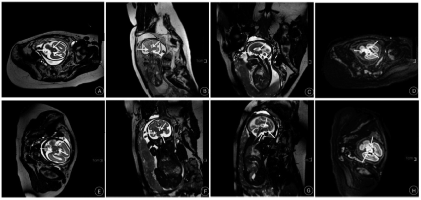
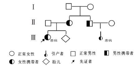
报道了1例胎儿颅内肿瘤病例。该例孕妇于孕24周四维超声提示胎儿颅内混合性回声,考虑颅内占位。我院超声复查亦显示胎儿颅内肿瘤;孕25周于我院行胎儿头磁共振成像(MRI)平扫示胎儿脑干前方肿瘤,邻近组织受压移位;孕29周第2次胎儿头MRI平扫示脑干前肿物较上次体积增大。于孕30+1周行引产手术。术后病理诊断符合MRI诊断。家系全外显子测序结果未发现致病基因变异,但检测到3个意义不明确的基因变异,其中ERBB2基因变异(参考变体:NM_001005862):c.3139C>T p.P1047S,源自其父亲。胎儿的姑姑曾生育一颅内肿瘤女孩,ERBB2基因也发生变异。数据库表明该基因变异的胶质瘤可能有家族聚集,与该病例相符。

关键词: 磁共振成像, 胎儿, 脑肿瘤, 神经胶质瘤, 超声检查, 产前诊断

Abstract:

We report a case of fetal cerebral tumor and family genetic analysis. In this case, the dimensional ultrasound showed the mixed intracranial echoes in the 24 weeks fetus. On the basis of that, the intracranial space-occupying lesion was considered. Ultrasound re-examination confirmed this consideration. The head magnetic resonance imaging (MRI) at 25 weeks of gestation age showed that the tumor were in the front of the fetal brainstem, and that the adjacent tissues were compressed and displaced. The second fetal MRI scan was done at 29 weeks of gestation age. This MRI showed that the tumor volume had increased. At 30+1 weeks, the mother of the fetus decided to have the option of labor induction. The postoperative pathological diagnosis was consistent with the MRI diagnosis. Whole exome sequencing did not find the pathogenic gene variation, but showed three gene variants with unclear significance. However, there was an interesting phenomenon. The ERBB2 gene variant c.3139C>T p.P1047Ss that originated from the father, was also detected in a cousin girl of the fetus in this family. Therfore, we considered that gliomas with ERBB2 gene mutation may be a family cluster.

Key words: Magnetic resonance imaging, Fetus, Brain neoplasms, Glioma, Ultrasonography, Prenatal diagnosis