| [1] |
Hamza A, Herr D, Solomayer EF, et al. Polyhydramnios: Causes, Diagnosis and Therapy[J]. Geburtshilfe Frauenheilkd, 2013, 73(12):1241-1246. doi: 10.1055/s-0033-1360163.
|
| [2] |
Huri M, Di Tommaso M, Seravalli V. Amniotic Fluid Disorders: From Prenatal Management to Neonatal Outcomes[J]. Children(Basel), 2023, 10(3):561. doi: 10.3390/children10030561.
|
| [3] |
Kouamé N, N′goan-Domoua AM, Nikiéma Z, et al. Polyhydramnios: a warning sign in the prenatal ultrasound diagnosis of foetal malformation?[J]. Diagn Interv Imaging, 2013, 94(4):433-437. doi: 10.1016/j.diii.2013.01.002.
|
| [4] |
Hwang DS, Mahdy H. Polyhydramnios[M]. StatPearls. Treasure Island (FL): StatPearls Publishing, 2023.
|
| [5] |
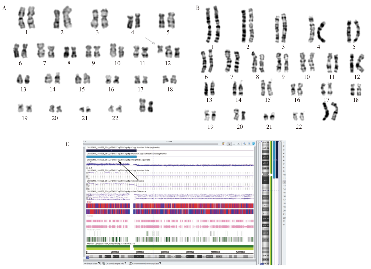
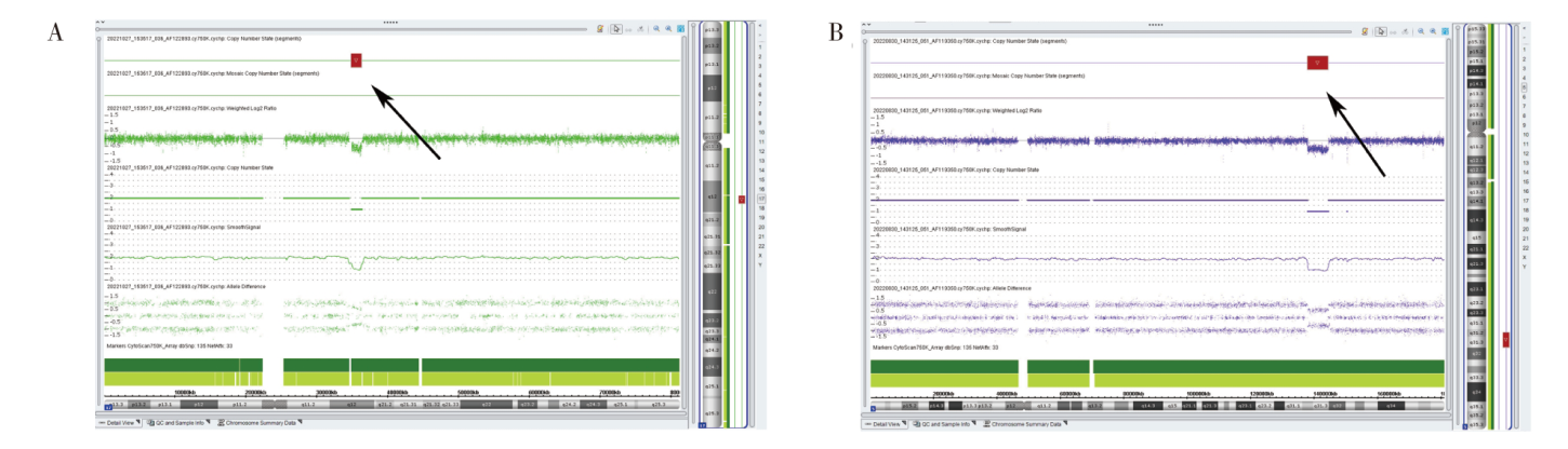
Qian G, Cai L, Yao H, et al. Chromosome microarray analysis combined with karyotype analysis is a powerful tool for the detection in pregnant women with high-risk indicators[J]. BMC Pregnancy Childbirth, 2023, 23(1):784. doi: 10.1186/s12884-023-06052-z.
|
| [6] |
Stosic M, Levy B, Wapner R. The Use of Chromosomal Microarray Analysis in Prenatal Diagnosis[J]. Obstet Gynecol Clin North Am, 2018, 45(1):55-68. doi: 10.1016/j.ogc.2017.10.002.
|
| [7] |
中国预防医学会出生缺陷预防与控制专业委员会产前筛查和诊断学组, 中华医学会医学遗传学分会产前诊断学组. 染色体微阵列分析技术在产前诊断中的应用指南(2023)[J]. 中华医学遗传学杂志, 2023, 40(9):1051-1061. doi: 10.3760/cma.j.cn112141-20230327-00146-1.
|
| [8] |
刘淑敏, 祁海云, 王生兰, 等. 孕妇羊水过多的原因及其与染色体异常的相关性[J]. 中华医学遗传学杂志, 2018, 35(4):607-608. doi: 10.3760/cma.j.issn.1003-9406.2018.04.033.
|
| [9] |
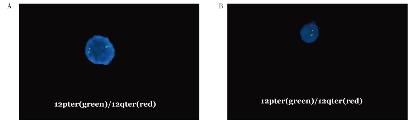
Zhuang J, Zhang N, Fu W, et al. Cytogenetic and molecular analysis of distal 4q duplication with distinctive phenotype using single-nucleotide polymorphism array[J]. Mol Cytogenet, 2021, 14(1):46. doi: 10.1186/s13039-021-00568-9.
pmid: 34587985
|
| [10] |
Riggs ER, Andersen EF, Cherry AM, et al. Technical standards for the interpretation and reporting of constitutional copy-number variants: a joint consensus recommendation of the American College of Medical Genetics and Genomics (ACMG) and the Clinical Genome Resource (ClinGen)[J]. Genet Med, 2020, 22(2):245-257. doi: 10.1038/s41436-019-0686-8.
pmid: 31690835
|
| [11] |
黄婧, 潘平山, 蒙达华, 等. 单核苷酸多态性微阵列芯片技术在羊水过多孕妇遗传学病因中的应用效果[J]. 广西医学, 2022, 44(15):1701-1704,1710. doi: 10.11675/j.issn.0253-4304.2022.15.03.
|
| [12] |
Shi P, Hou Y, Chen D, et al. Estimate of genetic variants using CNV-Seq for fetuses with oligohydramnios or polyhydramnios[J]. Mol Genet Genomic Med, 2023, 11(1):e2089. doi: 10.1002/mgg3.2089.
|
| [13] |
Kemeny S, Pebrel-Richard C, Gouas L, et al. Prenatal ultrasound diagnosis of a 48,XXYY syndrome[J]. Morphologie, 2013, 97(317):65-67. doi: 10.1016/j.morpho.2013.01.001.
pmid: 23473874
|
| [14] |
Fetta A, Toni F, Pettenuzzo I, et al. Structural brain abnormalities in Pallister-Killian syndrome: a neuroimaging study of 31 children[J]. Orphanet J Rare Dis, 2024, 19(1):107. doi: 10.1186/s13023-024-03065-5.
pmid: 38459574
|
| [15] |
方利元, 石晓梅, 刘倩, 等. 两例头围增大合并羊水过多的Pallister-Killian综合征胎儿的产前诊断[J]. 中华医学遗传学杂志, 2019, 36(3):278-280. doi: 10.3760/cma.j.issn.1003-9406.2019.03.022.
|
| [16] |
Mitchel MW, Moreno-De-Luca D, Myers SM, et al. 17q12 Recurrent Deletion Syndrome[M]. GeneReviews®. Seattle (WA): University of Washington, 2016.
|
| [17] |
黄丝琪, 张慧敏, 陈敏, 等. 染色体17q12微缺失或微重复综合征胎儿的超声特点及其遗传学和妊娠结局分析[J]. 中华妇产科杂志, 2023, 58(4):296-300. doi: 10.3760/cma.j.cn112141-20221110-00686.
|
| [18] |
Mautner VF, Kluwe L, Friedrich RE, et al. Clinical characterisation of 29 neurofibromatosis type-1 patients with molecularly ascertained 1.4 Mb type-1 NF1 deletions[J]. J Med Genet, 2010, 47(9):623-630. doi: 10.1136/jmg.2009.075937.
pmid: 20543202
|
 )
)