国际生殖健康/计划生育 ›› 2021, Vol. 40 ›› Issue (4): 265-271.doi: 10.12280/gjszjk.20210138
• 论著 • 下一篇
张月鑫, 刘菡文, 施陈楠, 宁松, 周静, 李楚玉, 杨晓玉, 覃莲菊, 刘嘉茵, 胡艳秋( ), 崔毓桂()
收稿日期:2021-03-22
出版日期:2021-07-15
发布日期:2021-07-27
通讯作者:
胡艳秋,崔毓桂
E-mail:huyanqiu78@163.com;cuiygnj@njmu.edu.cn
基金资助:
ZHANG Yue-xin, LIU Han-wen, SHI Chen-nan, NING Song, ZHOU Jing, LI Chu-yu, YANG Xiao-yu, QIN Lian-ju, LIU Jia-yin, HU Yan-qiu(), CUI Yu-gui()
Received:2021-03-22
Published:2021-07-15
Online:2021-07-27
Contact:
HU Yan-qiu,CUI Yu-gui
E-mail:huyanqiu78@163.com;cuiygnj@njmu.edu.cn
摘要:
目的: 探讨人羊膜间充质干细胞(hAMSC)改善高龄雄性小鼠睾丸功能的作用。方法: 以36周龄雄性小鼠为高龄模型组,10周龄小鼠为年轻对照组。31只高龄模型组小鼠随机分为5组,分别为生理盐水对照组、1%人血清白蛋白对照组和治疗组,其中生理盐水对照组3只,其余各组每组7只。治疗组小鼠尾静脉注射hAMSC,剂量分别为3.4×10⁶细胞/kg(低剂量组)、6.7×106细胞/kg(中剂量组)、1.4×107细胞/kg(高剂量组)。每周注射1次,治疗4次后小鼠继续饲养,5周后取血检测小鼠血清睾酮水平;行附睾内精子的分析;免疫荧光检测睾丸组织内STEM121和CD105的表达和定位,观察睾丸组织病理学改变,检测类固醇激素生成急性调节蛋白(StAR)、17β-羟基类固醇脱氢酶(17β-HSD)的表达水平。结果: 与年轻雄鼠比较,治疗前高龄雄鼠体质量增加、睾丸体质比下降、睾周脂肪质量增加(均P<0.01),血清睾酮水平下降(P<0.05),精子活动率降低、正常形态精子率下降(均P<0.05)。治疗前高龄雄鼠的精子计数、睾丸组织StAR和17β-HSD蛋白表达水平与年轻雄鼠比较差异无统计学意义(均P>0.05)。hAMSC治疗后,睾丸间质区可见STEM121和CD105表达的细胞;高剂量组血清睾酮水平升高(P<0.05);睾丸组织StAR蛋白表达无显著差异(P>0.05),但高剂量组17β-HSD蛋白表达上调(P<0.05)。中剂量组和高剂量组睾丸组织的生精小管细胞层数较人血清白蛋白对照组增多(P<0.05)。结论: 初步实验结果表明,hAMSC对睾丸衰老具有保护作用,并促进雄激素合成。
张月鑫, 刘菡文, 施陈楠, 宁松, 周静, 李楚玉, 杨晓玉, 覃莲菊, 刘嘉茵, 胡艳秋, 崔毓桂. 人羊膜间充质干细胞改善高龄小鼠睾丸功能的实验研究[J]. 国际生殖健康/计划生育, 2021, 40(4): 265-271.
ZHANG Yue-xin, LIU Han-wen, SHI Chen-nan, NING Song, ZHOU Jing, LI Chu-yu, YANG Xiao-yu, QIN Lian-ju, LIU Jia-yin, HU Yan-qiu, CUI Yu-gui. Human Amnion Mesenchymal Stem Cells Improve Testicular Steroidogenesis in Aging Mice[J]. Journal of International Reproductive Health/Family Planning, 2021, 40(4): 265-271.

图1 流式细胞术检测hAMSC表面分子标志物 注:CD44、CD73、CD90、CD105阳性率>95%,CD11b、CD19、CD34、CD45以及HLA-DR/DP/DQ阳性率<2%。
| 组别 | n | 体质量 (g) | 睾丸 体质比 | 睾周脂肪 质量(g) | 血清睾酮 (nmol/L) | StAR/ β-actin | 17β-HSD/ GAPDH | 精子计数 (个/mL,×106) | 精子活动率 (%) | 正常形态 精子率(%) | 生精小管 直径(μm) | 生精小管 细胞层数 |
|---|---|---|---|---|---|---|---|---|---|---|---|---|
| 年轻对照组 | 7 | 24.33±0.33 | 0.74±0.02 | 0.33±0.00 | 46.44±9.05 | 0.61±0.24 | 0.49±0.08 | 5.80±0.59 | 72.93±8.07 | 95.00±1.73 | 146.90±55.48 | 8.20±1.40 |
| 治疗前高龄组 | 7 | 37.67±0.88 | 0.57±0.02 | 2.42±0.27 | 12.91±2.25 | 0.65±0.31 | 0.44±0.18 | 5.88±1.58 | 52.57±9.25 | 85.43±2.76 | 190.90±27.07 | 5.80±1.22 |
| t | 14.14 | 4.86 | 7.61 | 3.60 | 0.23 | 0.51 | 0.08 | 2.87 | 5.09 | 3.92 | 7.09 | |
| P | 0.00 | 0.01 | 0.00 | 0.01 | 0.82 | 0.63 | 0.94 | 0.04 | 0.01 | 0.00 | 0.00 |
表1 hAMSC治疗前高龄组与年轻对照组比较 ($\bar{x} \pm s$)
| 组别 | n | 体质量 (g) | 睾丸 体质比 | 睾周脂肪 质量(g) | 血清睾酮 (nmol/L) | StAR/ β-actin | 17β-HSD/ GAPDH | 精子计数 (个/mL,×106) | 精子活动率 (%) | 正常形态 精子率(%) | 生精小管 直径(μm) | 生精小管 细胞层数 |
|---|---|---|---|---|---|---|---|---|---|---|---|---|
| 年轻对照组 | 7 | 24.33±0.33 | 0.74±0.02 | 0.33±0.00 | 46.44±9.05 | 0.61±0.24 | 0.49±0.08 | 5.80±0.59 | 72.93±8.07 | 95.00±1.73 | 146.90±55.48 | 8.20±1.40 |
| 治疗前高龄组 | 7 | 37.67±0.88 | 0.57±0.02 | 2.42±0.27 | 12.91±2.25 | 0.65±0.31 | 0.44±0.18 | 5.88±1.58 | 52.57±9.25 | 85.43±2.76 | 190.90±27.07 | 5.80±1.22 |
| t | 14.14 | 4.86 | 7.61 | 3.60 | 0.23 | 0.51 | 0.08 | 2.87 | 5.09 | 3.92 | 7.09 | |
| P | 0.00 | 0.01 | 0.00 | 0.01 | 0.82 | 0.63 | 0.94 | 0.04 | 0.01 | 0.00 | 0.00 |
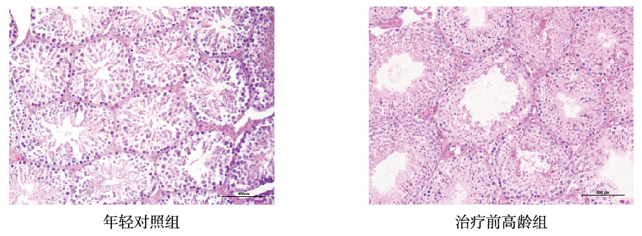
图2 治疗前高龄组与年轻对照组睾丸组织病理学形态图(HE染色x200) 注:高龄小鼠与年轻小鼠睾丸切片,标尺为100μm。

图3 hAMSC治疗 后高龄雄鼠睾丸间质区中有STEM121和CD105表达细胞 注:hAMSC治疗后高龄雄鼠睾丸组织免疫荧光图,绿色荧光为STEM121,红色荧光为CD105,标尺为50 μm。

图4 高龄雄鼠hAMSC治疗结束继续饲养5周后各组间体质量、睾丸体质比及睾周脂肪质量比较 注:1为生理盐水对照组;2为人血清白蛋白对照组;3为低剂量组;4为中剂量组;5为高剂量组。
| 组别 | n | 血清睾酮 | StAR/β-actin | 17β-HSD/GAPDH |
|---|---|---|---|---|
| 年轻对照组 | 7 | 46.44±9.05 | 0.61±0.10 | 0.49±0.04 |
| 治疗前高龄组 | 7 | 12.91±2.25 | 0.65±0.13 | 0.44±0.09 |
| 生理盐水对照组 | 3 | 16.87±3.31* | 0.70±0.21 | 0.51±0.11 |
| F | 9.79 | 0.13 | 0.27 | |
| P | 0.01 | 0.88 | 0.77 |
表2 生理盐水组和年轻对照及治疗前高龄组血清睾酮和睾酮合成相关酶比较 ($\bar{x} \pm s$)
| 组别 | n | 血清睾酮 | StAR/β-actin | 17β-HSD/GAPDH |
|---|---|---|---|---|
| 年轻对照组 | 7 | 46.44±9.05 | 0.61±0.10 | 0.49±0.04 |
| 治疗前高龄组 | 7 | 12.91±2.25 | 0.65±0.13 | 0.44±0.09 |
| 生理盐水对照组 | 3 | 16.87±3.31* | 0.70±0.21 | 0.51±0.11 |
| F | 9.79 | 0.13 | 0.27 | |
| P | 0.01 | 0.88 | 0.77 |
| 组别 | n | 血清睾酮 | StAR/β-actin | 17β-HSD/GAPDH |
|---|---|---|---|---|
| 生理盐水对照组 | 3 | 16.87±3.31 | 0.70±0.21 | 0.51±0.11 |
| 人血清白蛋白 对照组 | 7 | 14.85±2.72 | 0.54±0.18 | 0.49±0.11 |
| 低剂量组 | 7 | 18.36±3.41 | 0.49±0.19 | 0.48±0.19 |
| 中剂量组 | 7 | 15.24±1.92 | 0.55±0.17 | 0.71±0.10 |
| 高剂量组 | 7 | 28.51±9.40** | 0.84±0.33 | 0.82±0.09* |
| F | 5.56 | 1.63 | 4.53 | |
| P | 0.01 | 0.40 | 0.02 |
表3 hAMSC治疗后各组间血清睾酮和睾酮合成相关酶比较 ($\bar{x} \pm s$)
| 组别 | n | 血清睾酮 | StAR/β-actin | 17β-HSD/GAPDH |
|---|---|---|---|---|
| 生理盐水对照组 | 3 | 16.87±3.31 | 0.70±0.21 | 0.51±0.11 |
| 人血清白蛋白 对照组 | 7 | 14.85±2.72 | 0.54±0.18 | 0.49±0.11 |
| 低剂量组 | 7 | 18.36±3.41 | 0.49±0.19 | 0.48±0.19 |
| 中剂量组 | 7 | 15.24±1.92 | 0.55±0.17 | 0.71±0.10 |
| 高剂量组 | 7 | 28.51±9.40** | 0.84±0.33 | 0.82±0.09* |
| F | 5.56 | 1.63 | 4.53 | |
| P | 0.01 | 0.40 | 0.02 |
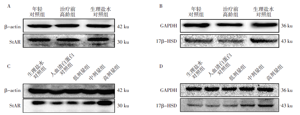
图5 hAMSC提高高龄雄鼠睾丸组织17β-HSD蛋白表达 注:A为StAR蛋白在年轻对照组、治疗前高龄组及生理盐水对照组高龄雄鼠睾丸中相对表达水平;B为17β-HSD蛋白在年轻对照组、治疗前高龄组及生理盐水对照组高龄雄鼠睾丸中相对表达水平;C为hAMSC治疗后,StAR蛋白在高龄雄鼠睾丸中相对表达水平;D为hAMSC治疗后,17β-HSD蛋白在高龄雄鼠睾丸中相对表达水平。
| 组别 | n | 精子计数(个/mL,×106) | 精子活动率(%) | 正常形态精子率(%) | 生精小管直径(μm) | 生精小管细胞层数 |
|---|---|---|---|---|---|---|
| 年轻对照组 | 7 | 5.80±0.59 | 72.93±8.07 | 95.00±1.73 | 146.92±55.48 | 8.20±1.40 |
| 治疗前高龄组 | 7 | 5.88±1.58 | 52.57±9.25 | 85.43±2.76 | 190.93±27.07 | 5.80±1.21 |
| 生理盐水对照组 | 3 | 7.04±1.14 | 54.87±19.09 | 78.60±0.66**# | 240.20±53.74**## | 7.00±1.74** |
| F | 0.97 | 2.17 | 55.31 | 29.59 | 20.03 | |
| P | 0.43 | 0.20 | 0.00 | 0.00 | 0.00 |
表4 生理盐水对照组、年轻对照组及治疗前高龄组精子参数及睾丸组织病理学比较 ($\bar{x} \pm s$)
| 组别 | n | 精子计数(个/mL,×106) | 精子活动率(%) | 正常形态精子率(%) | 生精小管直径(μm) | 生精小管细胞层数 |
|---|---|---|---|---|---|---|
| 年轻对照组 | 7 | 5.80±0.59 | 72.93±8.07 | 95.00±1.73 | 146.92±55.48 | 8.20±1.40 |
| 治疗前高龄组 | 7 | 5.88±1.58 | 52.57±9.25 | 85.43±2.76 | 190.93±27.07 | 5.80±1.21 |
| 生理盐水对照组 | 3 | 7.04±1.14 | 54.87±19.09 | 78.60±0.66**# | 240.20±53.74**## | 7.00±1.74** |
| F | 0.97 | 2.17 | 55.31 | 29.59 | 20.03 | |
| P | 0.43 | 0.20 | 0.00 | 0.00 | 0.00 |
| 组别 | n | 精子计数(个/mL,×106) | 精子活动率(%) | 正常形态精子率(%) | 生精小管直径(μm) | 生精小管细胞层数 |
|---|---|---|---|---|---|---|
| 生理盐水对照组 | 3 | 7.04±1.14 | 54.87±19.09 | 78.60±0.66 | 240.20±53.74 | 7.02±1.74 |
| 人血清白蛋白对照组 | 7 | 6.56±1.50 | 73.25±0.64 | 83.87±3.11 | 228.60±52.17 | 6.97±1.73 |
| 低剂量组 | 7 | 6.62±1.49 | 80.51±8.93 | 83.43±2.54 | 155.22±21.60** | 7.63±2.11 |
| 中剂量组 | 7 | 5.48±1.42 | 72.66±10.51 | 85.80±0.50 | 122.52±28.53** | 8.20±1.75** |
| 高剂量组 | 7 | 7.49±2.25 | 78.07±7.31 | 84.03±3.24 | 139.62±21.74** | 7.90±1.09* |
| F | 1.27 | 3.53 | 4.00 | 58.55 | 3.04 | |
| P | 0.32 | 0.02 | 0.03 | 0.00 | 0.02 |
表5 hAMSC治疗后各组间精子参数及睾丸组织病理学比较 ($\bar{x} \pm s$)
| 组别 | n | 精子计数(个/mL,×106) | 精子活动率(%) | 正常形态精子率(%) | 生精小管直径(μm) | 生精小管细胞层数 |
|---|---|---|---|---|---|---|
| 生理盐水对照组 | 3 | 7.04±1.14 | 54.87±19.09 | 78.60±0.66 | 240.20±53.74 | 7.02±1.74 |
| 人血清白蛋白对照组 | 7 | 6.56±1.50 | 73.25±0.64 | 83.87±3.11 | 228.60±52.17 | 6.97±1.73 |
| 低剂量组 | 7 | 6.62±1.49 | 80.51±8.93 | 83.43±2.54 | 155.22±21.60** | 7.63±2.11 |
| 中剂量组 | 7 | 5.48±1.42 | 72.66±10.51 | 85.80±0.50 | 122.52±28.53** | 8.20±1.75** |
| 高剂量组 | 7 | 7.49±2.25 | 78.07±7.31 | 84.03±3.24 | 139.62±21.74** | 7.90±1.09* |
| F | 1.27 | 3.53 | 4.00 | 58.55 | 3.04 | |
| P | 0.32 | 0.02 | 0.03 | 0.00 | 0.02 |
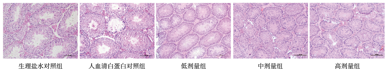
图6 hAMSC治疗后睾丸组织病理学形态图( HE染色x200) 注:hAMSC治疗后高龄小鼠睾丸切片,标尺为100 μm。
| [1] |
朱伟杰. 高龄男性生育研究的机遇与挑战[J]. 中华生殖与避孕杂志, 2019, 39(6):433-435. doi: 10.3760/cma.j.issn.2096-2916.2019. 06.001.
doi: 10.3760/cma.j.issn.2096-2916.2019. 06.001 |
| [2] |
Santiago J, Silva JV, Alves MG, et al. Testicular Aging: An Overview of Ultrastructural, Cellular, and Molecular Alterations[J]. J Gerontol A Biol Sci Med Sci, 2019, 74(6):860-871. doi: 10.1093/gerona/gly082.
doi: 10.1093/gerona/gly082 pmid: 29688289 |
| [3] |
Mann U, Shiff B, Patel P. Reasons for worldwide decline in male fertility[J]. Curr Opin Urol, 2020, 30(3):296-301. doi: 10.1097/MOU.0000000000000745.
doi: 10.1097/MOU.0000000000000745 URL |
| [4] |
Gagliano-Jucá T, Basaria S. Testosterone replacement therapy and cardiovascular risk[J]. Nat Rev Cardiol, 2019, 16(9):555-574. doi: 10.1038/s41569-019-0211-4.
doi: 10.1038/s41569-019-0211-4 pmid: 31123340 |
| [5] |
Bhasin S. Testosterone replacement in aging men: an evidence-based patient-centric perspective[J]. J Clin Invest, 2021, 131(4):e146607. doi: 10.1172/JCI146607.
doi: 10.1172/JCI146607 URL |
| [6] |
Halvaei I, Litzky J, Esfandiari N. Advanced paternal age: effects on sperm parameters, assisted reproduction outcomes and offspring health[J]. Reprod Biol Endocrinol, 2020, 18(1):110. doi: 10.1186/s12958-020-00668-y.
doi: 10.1186/s12958-020-00668-y URL |
| [7] |
刘菡文, 覃莲菊, 崔毓桂. 间充质干细胞分泌因子调节氧化应激的作用[J]. 国际生殖健康/计划生育杂志, 2019, 38(6):493-497. doi: 10.3969/j.issn.1674-1889.2019.06.013.
doi: 10.3969/j.issn.1674-1889.2019.06.013 |
| [8] |
钱孝鑫, 刘艳. 间充质干细胞治疗男性不育的研究进展[J]. 中华男科学杂志, 2020, 26(6):564-569. doi: 10.13263/j.cnki.nja.2020.06.014.
doi: 10.13263/j.cnki.nja.2020.06.014 |
| [9] |
张琴静, 陈爱琴, 宁松, 等. 建立不同类型干细胞并比较其分泌细胞因子的水平[J]. 生殖医学杂志, 2016, 25(6):528-539. doi: 10.3969/j.issn.1004-3845.2016.06.009.
doi: 10.3969/j.issn.1004-3845.2016.06.009 |
| [10] |
Naji A, Eitoku M, Favier B, et al. Biological functions of mesenchymal stem cells and clinical implications[J]. Cell Mol Life Sci, 2019, 76(17):3323-3348. doi: 10.1007/s00018-019-03125-1.
doi: 10.1007/s00018-019-03125-1 URL |
| [11] |
Ding C, Zou Q, Wang F, et al. Human amniotic mesenchymal stem cells improve ovarian function in natural aging through secreting hepatocyte growth factor and epidermal growth factor[J]. Stem Cell Res Ther, 2018, 9(1):55. doi: 10.1186/s13287-018-0781-9.
doi: 10.1186/s13287-018-0781-9 URL |
| [12] | 蒋春艳. 人羊膜间充质干细胞修复卵巢功能的实验研究[D]. 南京:南京医科大学, 2014. |
| [13] |
Jannini EA, Nappi RE. Couplepause: A New Paradigm in Treating Sexual Dysfunction During Menopause and Andropause[J]. Sex Med Rev, 2018, 6(3):384-395. doi: 10.1016/j.sxmr.2017.11.002.
doi: 10.1016/j.sxmr.2017.11.002 |
| [14] |
Miller WL. MECHANISMS IN ENDOCRINOLOGY: Rare defects in adrenal steroidogenesis[J]. Eur J Endocrinol, 2018, 179(3):R125-R141. doi: 10.1530/EJE-18-0279.
doi: 10.1530/EJE-18-0279 URL |
| [15] |
Yu SJ, Wang YC, Chang CY, et al. NanoCsA improves the survival of human iPSC transplant in hemiparkinsonian rats[J]. Brain Res, 2019, 1719:124-132. doi: 10.1016/j.brainres.2019.05.040.
doi: 10.1016/j.brainres.2019.05.040 URL |
| [16] |
Brown C, McKee C, Bakshi S, et al. Mesenchymal stem cells: Cell therapy and regeneration potential[J]. J Tissue Eng Regen Med, 2019, 13(9):1738-1755. doi: 10.1002/term.2914.
doi: 10.1002/term.2914 URL |
| [17] |
Qian C, Meng Q, Lu J, et al. Human amnion mesenchymal stem cells restore spermatogenesis in mice with busulfan-induced testis toxicity by inhibiting apoptosis and oxidative stress[J]. Stem Cell Res Ther, 2020, 11(1):290. doi: 10.1186/s13287-020-01803-7.
doi: 10.1186/s13287-020-01803-7 URL |
| [18] |
Chen H, Tang QL, Wu XY, et al. Differentiation of human umbilical cord mesenchymal stem cells into germ-like cells in mouse seminiferous tubules[J]. Mol Med Rep, 2015, 12(1):819-828. doi: 10.3892/mmr.2015.3528.
doi: 10.3892/mmr.2015.3528 pmid: 25815600 |
| [19] |
Zirkin BR, Papadopoulos V. Leydig cells: formation, function, and regulation[J]. Biol Reprod, 2018, 99(1):101-111. doi: 10.1093/biolre/ioy059.
doi: 10.1093/biolre/ioy059 pmid: 29566165 |
| [20] | 张琴静. 干细胞分泌因子对卵巢功能减退的治疗作用及机制研究[D]. 南京:南京医科大学, 2016. |
| [21] |
Khamis T, Abdelalim AF, Abdallah SH, et al. Early intervention with breast milk mesenchymal stem cells attenuates the development of diabetic-induced testicular dysfunction via hypothalamic Kisspeptin/Kiss1r-GnRH/GnIH system in male rats[J]. Biochim Biophys Acta Mol Basis Dis, 2020, 1866(1):165577. doi: 10.1016/j.bbadis.2019.165577.
doi: 10.1016/j.bbadis.2019.165577 URL |
| [1] | 吴静, 刘聪, 谢青贞. 微塑料暴露对雌性及其子代健康的影响[J]. 国际生殖健康/计划生育杂志, 2024, 43(2): 155-158. |
| [2] | 叶明珠, 郑洁, 李杰芃, 许莉欣. 医源性卵巢储备功能减退患者的卵母细胞冷冻生育力保存应用[J]. 国际生殖健康/计划生育杂志, 2023, 42(6): 498-502. |
| [3] | 邓美香, 石一柱, 冯兰青. 内分泌干扰物对女性生育力和辅助生殖技术结局的影响[J]. 国际生殖健康/计划生育杂志, 2023, 42(4): 304-309. |
| [4] | 李延, 胡方方, 陈欢欢, 张磊, 张翠莲, 梁琳琳. 窦前卵泡体外三维培养系统研究进展[J]. 国际生殖健康/计划生育杂志, 2023, 42(3): 221-225. |
| [5] | 张丽洪, 胡荣, 谢雷, 许晓娟, 胡俊平. 新型冠状病毒感染对男性精液检测指标的影响[J]. 国际生殖健康/计划生育杂志, 2023, 42(3): 231-235. |
| [6] | 杨志娟, 姚婷, 侯海燕. 线粒体自噬与卵巢功能[J]. 国际生殖健康/计划生育杂志, 2023, 42(3): 240-244. |
| [7] | 曾中虹, 梁婷. 不同膳食模式对女性生育力的影响[J]. 国际生殖健康/计划生育杂志, 2023, 42(3): 250-253. |
| [8] | 叶玲, 马雪影, 王晓慧. SARS-CoV-2对生育的影响[J]. 国际生殖健康/计划生育, 2022, 41(6): 482-486. |
| [9] | 李姗姗, 申永梅, 卫卓, 陈凌, 姚立英, 张蕾, 李雯, 曹家松, 常颖. 胎盘嵌合型16三体合并胎儿生长受限一例[J]. 国际生殖健康/计划生育, 2022, 41(3): 207-209. |
| [10] | 王青娣, 熊正方. 维生素D对辅助生殖技术助孕结局的影响[J]. 国际生殖健康/计划生育, 2022, 41(2): 143-146. |
| [11] | 杜洁冰, 杨一华. 早发性卵巢功能不全患者生育力改善策略的研究进展[J]. 国际生殖健康/计划生育, 2021, 40(5): 397-401. |
| [12] | 戴垒, 冒韵东. 子宫内膜干/祖细胞与子宫内膜异位症发病机制的研究进展[J]. 国际生殖健康/计划生育, 2021, 40(4): 319-322. |
| [13] | 叶晨霞, 应小燕. 间充质干细胞治疗宫腔粘连的研究进展[J]. 国际生殖健康/计划生育, 2021, 40(3): 242-246. |
| [14] | 袁静, 陈超, 张颖. 慢性子宫内膜炎对生育影响的研究进展[J]. 国际生殖健康/计划生育, 2021, 40(3): 256-259. |
| [15] | 程铭, 贾婵维, 刘英. 早发性卵巢功能不全的临床诊疗进展[J]. 国际生殖健康/计划生育, 2021, 40(2): 137-141. |
| 阅读次数 | ||||||
|
全文 |
|
|||||
|
摘要 |
|
|||||