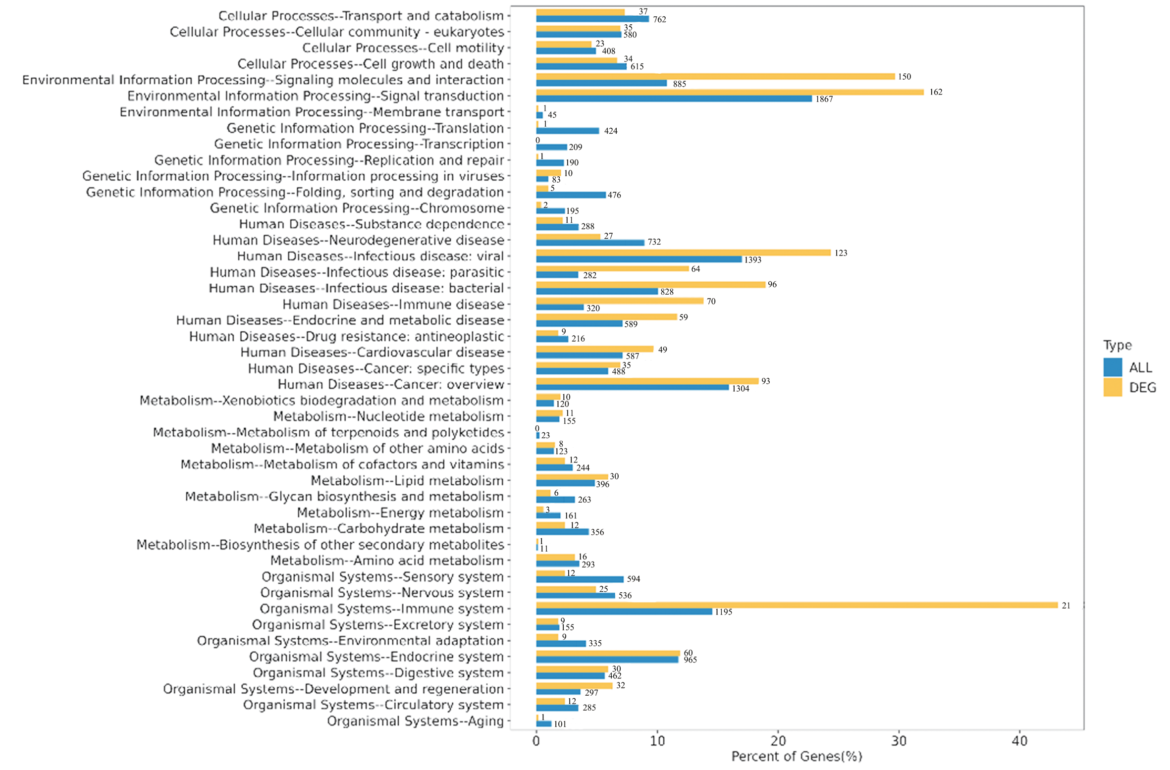
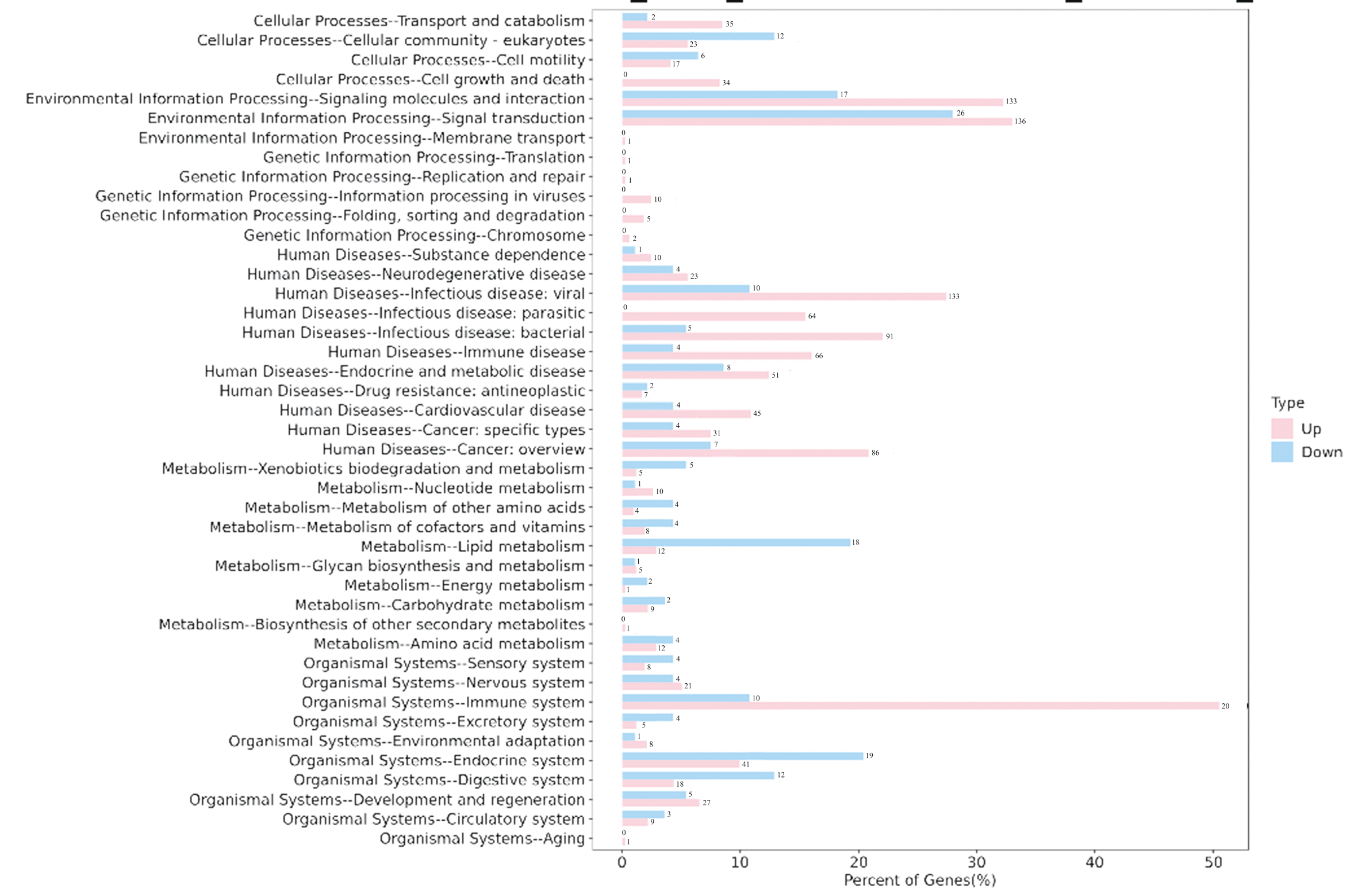
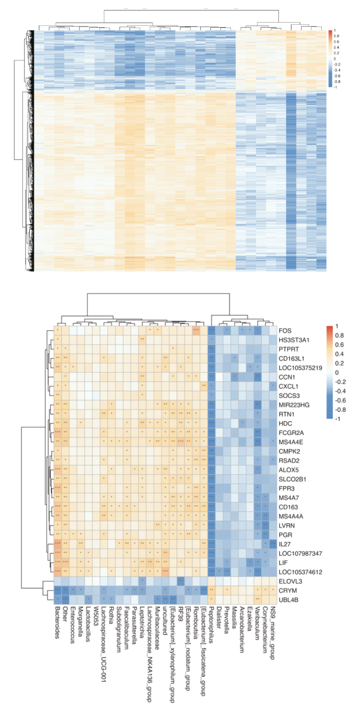
| [1] |
Gaines T, Simhan J. Adult Hypospadias Outcomes for the Pediatric Urologist[J]. Curr Urol Rep, 2024, 25(4):63-70. doi: 10.1007/s11934-024-01196-7.
|
| [2] |
唐耘熳, 刘愉, 王学军, 等. 尿道下裂初次矫治手术主要操作对阴茎长度的影响研究[J]. 中国修复重建外科杂志, 2022, 36(2):231-235. doi: 10.7507/1002-1892.202109038.
|
| [3] |
Lederberg J. Infectious history[J]. Science, 2000, 288(5464):287-293. doi: 10.1126/science.288.5464.287.
pmid: 10777411
|
| [4] |
Colella M, Topi S, Palmirotta R, et al. An Overview of the Microbiota of the Human Urinary Tract in Health and Disease: Current Issues and Perspectives[J]. Life (Basel), 2023, 13(7):1486. doi: 10.3390/life13071486.
|
| [5] |
Jamil ML, Perecman A, Sherman A, et al. Urinary microbiome differences between lichen sclerosus induced and non-lichen sclerosus induced urethral stricture disease[J]. World J Urol, 2023, 41(9):2495-2501. doi: 10.1007/s00345-023-04490-0.
pmid: 37421420
|
| [6] |
Isali I, Wong TR, Wu CW, et al. Genomic Risk Factors for Urethral Stricture: A Systematic Review and Gene Network Analysis[J]. Urology, 2024, 184:251-258. doi: 10.1016/j.urology.2023.12.014.
|
| [7] |
Ma Y, Liu Z, Miao L, et al. Mechanisms underlying pathological scarring by fibroblasts during wound healing[J]. Int Wound J, 2023, 20(6):2190-2206. doi: 10.1111/iwj.14097.
pmid: 36726192
|
| [8] |
Werneburg GT, Hsieh MH. Clinical Microbiome Testing for Urology[J]. Urol Clin North Am, 2024, 51(4):493-504. doi: 10.1016/j.ucl.2024.06.007.
|
| [9] |
Choi HW, Lee KW, Kim YH. Microbiome in urological diseases: Axis crosstalk and bladder disorders[J]. Investig Clin Urol, 2023, 64(2):126-139. doi: 10.4111/icu.20220357.
pmid: 36882171
|
| [10] |
Zhao Z, Cheng W, Qu W, et al. Antibiotic Alleviates Radiation-Induced Intestinal Injury by Remodeling Microbiota, Reducing Inflammation, and Inhibiting Fibrosis[J]. ACS Omega, 2020, 5(6):2967-2977. doi: 10.1021/acsomega.9b03906.
pmid: 32095719
|
| [11] |
Zhou Y, Zhou Z, Zheng L, et al. Urinary Tract Infections Caused by Uropathogenic Escherichia coli: Mechanisms of Infection and Treatment Options[J]. Int J Mol Sci, 2023, 24(13):10537. doi: 10.3390/ijms241310537.
|
| [12] |
Hreha TN, Collins CA, Daugherty AL, et al. TGFβ1 orchestrates renal fibrosis following Escherichia coli pyelonephritis[J]. Physiol Rep, 2020, 8(6):e14401. doi: 10.14814/phy2.14401.
|
| [13] |
Ong CH, Tham CL, Harith HH, et al. TGF-β-induced fibrosis: A review on the underlying mechanism and potential therapeutic strategies[J]. Eur J Pharmacol, 2021, 911:174510. doi: 10.1016/j.ejphar.2021.174510.
|
| [14] |
Verma BK, Chatterjee A, Kondaiah P, et al. Substrate Stiffness Modulates TGF-β Activation and ECM-Associated Gene Expression in Fibroblasts[J]. Bioengineering(Basel), 2023, 10(9):998. doi: 10.3390/bioengineering10090998.
|
 )
)