Journal of International Reproductive Health/Family Planning ›› 2021, Vol. 40 ›› Issue (4): 265-271.doi: 10.12280/gjszjk.20210138
• Original Article • Next Articles
ZHANG Yue-xin, LIU Han-wen, SHI Chen-nan, NING Song, ZHOU Jing, LI Chu-yu, YANG Xiao-yu, QIN Lian-ju, LIU Jia-yin, HU Yan-qiu( ), CUI Yu-gui()
Received:2021-03-22
Published:2021-07-15
Online:2021-07-27
Contact:
HU Yan-qiu,CUI Yu-gui
E-mail:huyanqiu78@163.com;cuiygnj@njmu.edu.cn
ZHANG Yue-xin, LIU Han-wen, SHI Chen-nan, NING Song, ZHOU Jing, LI Chu-yu, YANG Xiao-yu, QIN Lian-ju, LIU Jia-yin, HU Yan-qiu, CUI Yu-gui. Human Amnion Mesenchymal Stem Cells Improve Testicular Steroidogenesis in Aging Mice[J]. Journal of International Reproductive Health/Family Planning, 2021, 40(4): 265-271.
Add to citation manager EndNote|Ris|BibTeX

| 组别 | n | 体质量 (g) | 睾丸 体质比 | 睾周脂肪 质量(g) | 血清睾酮 (nmol/L) | StAR/ β-actin | 17β-HSD/ GAPDH | 精子计数 (个/mL,×106) | 精子活动率 (%) | 正常形态 精子率(%) | 生精小管 直径(μm) | 生精小管 细胞层数 |
|---|---|---|---|---|---|---|---|---|---|---|---|---|
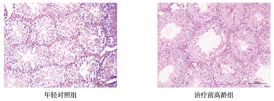
| 年轻对照组 | 7 | 24.33±0.33 | 0.74±0.02 | 0.33±0.00 | 46.44±9.05 | 0.61±0.24 | 0.49±0.08 | 5.80±0.59 | 72.93±8.07 | 95.00±1.73 | 146.90±55.48 | 8.20±1.40 |
| 治疗前高龄组 | 7 | 37.67±0.88 | 0.57±0.02 | 2.42±0.27 | 12.91±2.25 | 0.65±0.31 | 0.44±0.18 | 5.88±1.58 | 52.57±9.25 | 85.43±2.76 | 190.90±27.07 | 5.80±1.22 |
| t | 14.14 | 4.86 | 7.61 | 3.60 | 0.23 | 0.51 | 0.08 | 2.87 | 5.09 | 3.92 | 7.09 | |
| P | 0.00 | 0.01 | 0.00 | 0.01 | 0.82 | 0.63 | 0.94 | 0.04 | 0.01 | 0.00 | 0.00 |
| 组别 | n | 体质量 (g) | 睾丸 体质比 | 睾周脂肪 质量(g) | 血清睾酮 (nmol/L) | StAR/ β-actin | 17β-HSD/ GAPDH | 精子计数 (个/mL,×106) | 精子活动率 (%) | 正常形态 精子率(%) | 生精小管 直径(μm) | 生精小管 细胞层数 |
|---|---|---|---|---|---|---|---|---|---|---|---|---|
| 年轻对照组 | 7 | 24.33±0.33 | 0.74±0.02 | 0.33±0.00 | 46.44±9.05 | 0.61±0.24 | 0.49±0.08 | 5.80±0.59 | 72.93±8.07 | 95.00±1.73 | 146.90±55.48 | 8.20±1.40 |
| 治疗前高龄组 | 7 | 37.67±0.88 | 0.57±0.02 | 2.42±0.27 | 12.91±2.25 | 0.65±0.31 | 0.44±0.18 | 5.88±1.58 | 52.57±9.25 | 85.43±2.76 | 190.90±27.07 | 5.80±1.22 |
| t | 14.14 | 4.86 | 7.61 | 3.60 | 0.23 | 0.51 | 0.08 | 2.87 | 5.09 | 3.92 | 7.09 | |
| P | 0.00 | 0.01 | 0.00 | 0.01 | 0.82 | 0.63 | 0.94 | 0.04 | 0.01 | 0.00 | 0.00 |


| 组别 | n | 血清睾酮 | StAR/β-actin | 17β-HSD/GAPDH |
|---|---|---|---|---|
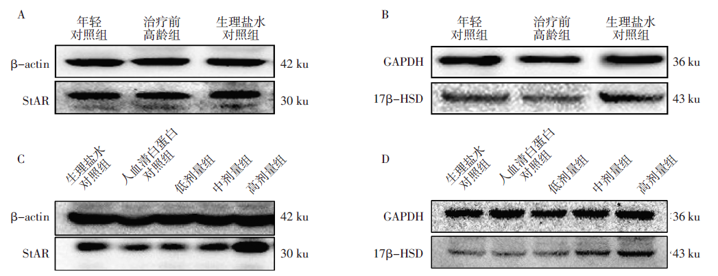
| 年轻对照组 | 7 | 46.44±9.05 | 0.61±0.10 | 0.49±0.04 |
| 治疗前高龄组 | 7 | 12.91±2.25 | 0.65±0.13 | 0.44±0.09 |
| 生理盐水对照组 | 3 | 16.87±3.31* | 0.70±0.21 | 0.51±0.11 |
| F | 9.79 | 0.13 | 0.27 | |
| P | 0.01 | 0.88 | 0.77 |
| 组别 | n | 血清睾酮 | StAR/β-actin | 17β-HSD/GAPDH |
|---|---|---|---|---|
| 年轻对照组 | 7 | 46.44±9.05 | 0.61±0.10 | 0.49±0.04 |
| 治疗前高龄组 | 7 | 12.91±2.25 | 0.65±0.13 | 0.44±0.09 |
| 生理盐水对照组 | 3 | 16.87±3.31* | 0.70±0.21 | 0.51±0.11 |
| F | 9.79 | 0.13 | 0.27 | |
| P | 0.01 | 0.88 | 0.77 |
| 组别 | n | 血清睾酮 | StAR/β-actin | 17β-HSD/GAPDH |
|---|---|---|---|---|
| 生理盐水对照组 | 3 | 16.87±3.31 | 0.70±0.21 | 0.51±0.11 |
| 人血清白蛋白 对照组 | 7 | 14.85±2.72 | 0.54±0.18 | 0.49±0.11 |
| 低剂量组 | 7 | 18.36±3.41 | 0.49±0.19 | 0.48±0.19 |
| 中剂量组 | 7 | 15.24±1.92 | 0.55±0.17 | 0.71±0.10 |
| 高剂量组 | 7 | 28.51±9.40** | 0.84±0.33 | 0.82±0.09* |
| F | 5.56 | 1.63 | 4.53 | |
| P | 0.01 | 0.40 | 0.02 |
| 组别 | n | 精子计数(个/mL,×106) | 精子活动率(%) | 正常形态精子率(%) | 生精小管直径(μm) | 生精小管细胞层数 |
|---|---|---|---|---|---|---|
| 年轻对照组 | 7 | 5.80±0.59 | 72.93±8.07 | 95.00±1.73 | 146.92±55.48 | 8.20±1.40 |
| 治疗前高龄组 | 7 | 5.88±1.58 | 52.57±9.25 | 85.43±2.76 | 190.93±27.07 | 5.80±1.21 |
| 生理盐水对照组 | 3 | 7.04±1.14 | 54.87±19.09 | 78.60±0.66**# | 240.20±53.74**## | 7.00±1.74** |
| F | 0.97 | 2.17 | 55.31 | 29.59 | 20.03 | |
| P | 0.43 | 0.20 | 0.00 | 0.00 | 0.00 |
| 组别 | n | 精子计数(个/mL,×106) | 精子活动率(%) | 正常形态精子率(%) | 生精小管直径(μm) | 生精小管细胞层数 |
|---|---|---|---|---|---|---|
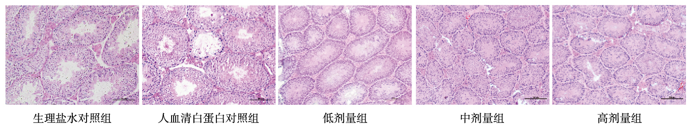
| 生理盐水对照组 | 3 | 7.04±1.14 | 54.87±19.09 | 78.60±0.66 | 240.20±53.74 | 7.02±1.74 |
| 人血清白蛋白对照组 | 7 | 6.56±1.50 | 73.25±0.64 | 83.87±3.11 | 228.60±52.17 | 6.97±1.73 |
| 低剂量组 | 7 | 6.62±1.49 | 80.51±8.93 | 83.43±2.54 | 155.22±21.60** | 7.63±2.11 |
| 中剂量组 | 7 | 5.48±1.42 | 72.66±10.51 | 85.80±0.50 | 122.52±28.53** | 8.20±1.75** |
| 高剂量组 | 7 | 7.49±2.25 | 78.07±7.31 | 84.03±3.24 | 139.62±21.74** | 7.90±1.09* |
| F | 1.27 | 3.53 | 4.00 | 58.55 | 3.04 | |
| P | 0.32 | 0.02 | 0.03 | 0.00 | 0.02 |
| 组别 | n | 精子计数(个/mL,×106) | 精子活动率(%) | 正常形态精子率(%) | 生精小管直径(μm) | 生精小管细胞层数 |
|---|---|---|---|---|---|---|
| 生理盐水对照组 | 3 | 7.04±1.14 | 54.87±19.09 | 78.60±0.66 | 240.20±53.74 | 7.02±1.74 |
| 人血清白蛋白对照组 | 7 | 6.56±1.50 | 73.25±0.64 | 83.87±3.11 | 228.60±52.17 | 6.97±1.73 |
| 低剂量组 | 7 | 6.62±1.49 | 80.51±8.93 | 83.43±2.54 | 155.22±21.60** | 7.63±2.11 |
| 中剂量组 | 7 | 5.48±1.42 | 72.66±10.51 | 85.80±0.50 | 122.52±28.53** | 8.20±1.75** |
| 高剂量组 | 7 | 7.49±2.25 | 78.07±7.31 | 84.03±3.24 | 139.62±21.74** | 7.90±1.09* |
| F | 1.27 | 3.53 | 4.00 | 58.55 | 3.04 | |
| P | 0.32 | 0.02 | 0.03 | 0.00 | 0.02 |
| [1] |
朱伟杰. 高龄男性生育研究的机遇与挑战[J]. 中华生殖与避孕杂志, 2019, 39(6):433-435. doi: 10.3760/cma.j.issn.2096-2916.2019. 06.001.
doi: 10.3760/cma.j.issn.2096-2916.2019. 06.001 |
| [2] |
Santiago J, Silva JV, Alves MG, et al. Testicular Aging: An Overview of Ultrastructural, Cellular, and Molecular Alterations[J]. J Gerontol A Biol Sci Med Sci, 2019, 74(6):860-871. doi: 10.1093/gerona/gly082.
doi: 10.1093/gerona/gly082 pmid: 29688289 |
| [3] |
Mann U, Shiff B, Patel P. Reasons for worldwide decline in male fertility[J]. Curr Opin Urol, 2020, 30(3):296-301. doi: 10.1097/MOU.0000000000000745.
doi: 10.1097/MOU.0000000000000745 URL |
| [4] |
Gagliano-Jucá T, Basaria S. Testosterone replacement therapy and cardiovascular risk[J]. Nat Rev Cardiol, 2019, 16(9):555-574. doi: 10.1038/s41569-019-0211-4.
doi: 10.1038/s41569-019-0211-4 pmid: 31123340 |
| [5] |
Bhasin S. Testosterone replacement in aging men: an evidence-based patient-centric perspective[J]. J Clin Invest, 2021, 131(4):e146607. doi: 10.1172/JCI146607.
doi: 10.1172/JCI146607 URL |
| [6] |
Halvaei I, Litzky J, Esfandiari N. Advanced paternal age: effects on sperm parameters, assisted reproduction outcomes and offspring health[J]. Reprod Biol Endocrinol, 2020, 18(1):110. doi: 10.1186/s12958-020-00668-y.
doi: 10.1186/s12958-020-00668-y URL |
| [7] |
刘菡文, 覃莲菊, 崔毓桂. 间充质干细胞分泌因子调节氧化应激的作用[J]. 国际生殖健康/计划生育杂志, 2019, 38(6):493-497. doi: 10.3969/j.issn.1674-1889.2019.06.013.
doi: 10.3969/j.issn.1674-1889.2019.06.013 |
| [8] |
钱孝鑫, 刘艳. 间充质干细胞治疗男性不育的研究进展[J]. 中华男科学杂志, 2020, 26(6):564-569. doi: 10.13263/j.cnki.nja.2020.06.014.
doi: 10.13263/j.cnki.nja.2020.06.014 |
| [9] |
张琴静, 陈爱琴, 宁松, 等. 建立不同类型干细胞并比较其分泌细胞因子的水平[J]. 生殖医学杂志, 2016, 25(6):528-539. doi: 10.3969/j.issn.1004-3845.2016.06.009.
doi: 10.3969/j.issn.1004-3845.2016.06.009 |
| [10] |
Naji A, Eitoku M, Favier B, et al. Biological functions of mesenchymal stem cells and clinical implications[J]. Cell Mol Life Sci, 2019, 76(17):3323-3348. doi: 10.1007/s00018-019-03125-1.
doi: 10.1007/s00018-019-03125-1 URL |
| [11] |
Ding C, Zou Q, Wang F, et al. Human amniotic mesenchymal stem cells improve ovarian function in natural aging through secreting hepatocyte growth factor and epidermal growth factor[J]. Stem Cell Res Ther, 2018, 9(1):55. doi: 10.1186/s13287-018-0781-9.
doi: 10.1186/s13287-018-0781-9 URL |
| [12] | 蒋春艳. 人羊膜间充质干细胞修复卵巢功能的实验研究[D]. 南京:南京医科大学, 2014. |
| [13] |
Jannini EA, Nappi RE. Couplepause: A New Paradigm in Treating Sexual Dysfunction During Menopause and Andropause[J]. Sex Med Rev, 2018, 6(3):384-395. doi: 10.1016/j.sxmr.2017.11.002.
doi: 10.1016/j.sxmr.2017.11.002 |
| [14] |
Miller WL. MECHANISMS IN ENDOCRINOLOGY: Rare defects in adrenal steroidogenesis[J]. Eur J Endocrinol, 2018, 179(3):R125-R141. doi: 10.1530/EJE-18-0279.
doi: 10.1530/EJE-18-0279 URL |
| [15] |
Yu SJ, Wang YC, Chang CY, et al. NanoCsA improves the survival of human iPSC transplant in hemiparkinsonian rats[J]. Brain Res, 2019, 1719:124-132. doi: 10.1016/j.brainres.2019.05.040.
doi: 10.1016/j.brainres.2019.05.040 URL |
| [16] |
Brown C, McKee C, Bakshi S, et al. Mesenchymal stem cells: Cell therapy and regeneration potential[J]. J Tissue Eng Regen Med, 2019, 13(9):1738-1755. doi: 10.1002/term.2914.
doi: 10.1002/term.2914 URL |
| [17] |
Qian C, Meng Q, Lu J, et al. Human amnion mesenchymal stem cells restore spermatogenesis in mice with busulfan-induced testis toxicity by inhibiting apoptosis and oxidative stress[J]. Stem Cell Res Ther, 2020, 11(1):290. doi: 10.1186/s13287-020-01803-7.
doi: 10.1186/s13287-020-01803-7 URL |
| [18] |
Chen H, Tang QL, Wu XY, et al. Differentiation of human umbilical cord mesenchymal stem cells into germ-like cells in mouse seminiferous tubules[J]. Mol Med Rep, 2015, 12(1):819-828. doi: 10.3892/mmr.2015.3528.
doi: 10.3892/mmr.2015.3528 pmid: 25815600 |
| [19] |
Zirkin BR, Papadopoulos V. Leydig cells: formation, function, and regulation[J]. Biol Reprod, 2018, 99(1):101-111. doi: 10.1093/biolre/ioy059.
doi: 10.1093/biolre/ioy059 pmid: 29566165 |
| [20] | 张琴静. 干细胞分泌因子对卵巢功能减退的治疗作用及机制研究[D]. 南京:南京医科大学, 2016. |
| [21] |
Khamis T, Abdelalim AF, Abdallah SH, et al. Early intervention with breast milk mesenchymal stem cells attenuates the development of diabetic-induced testicular dysfunction via hypothalamic Kisspeptin/Kiss1r-GnRH/GnIH system in male rats[J]. Biochim Biophys Acta Mol Basis Dis, 2020, 1866(1):165577. doi: 10.1016/j.bbadis.2019.165577.
doi: 10.1016/j.bbadis.2019.165577 URL |
| [1] | YANG Qin, WANG Han-ting, CAO Yuan-yuan, ZHOU Jun, WANG Gui-ling. Effect of Resveratrol on the Function of Ovarian Granulose Cells [J]. Journal of International Reproductive Health/Family Planning, 2024, 43(6): 524-528. |
| [2] | LI Xuan-ang, WANG Ting-ting, XIANG Shan, ZHAO Shuai, LIAN Fang. Research Progress of Ferroptosis in Pathogenesis of Polycystic Ovary Syndrome [J]. Journal of International Reproductive Health/Family Planning, 2024, 43(5): 425-429. |
| [3] | ZHANG Ai-yu, LUAN Cui-yu, WANG Dong-mei, JIANG Shuai. Analysis on the Status Quo and Influencing Factors of Medical Treatment Delay in Infertility Patients Undergoing IVF-ET [J]. Journal of International Reproductive Health/Family Planning, 2024, 43(3): 190-194. |
| [4] | LIU Shu-jie, LI Ming-ze, ZHANG Hai-yan. Modium-Low Differentiation Sertoli-Leydig Cell Tumor of the Ovary: A Case Report and Literature Review [J]. Journal of International Reproductive Health/Family Planning, 2024, 43(3): 207-211. |
| [5] | HE Qing-wen, LI Xi-hong. Research Progress on Sleep Disorders in Patients Receiving Assisted Reproductive Technology and Non-Pharmacological Intervention [J]. Journal of International Reproductive Health/Family Planning, 2024, 43(3): 234-237. |
| [6] | GAO Zhao-yang, ZHANG Ning-qing, CHEN Qiong-hua, WU Rong-feng. The Role of CircRNAs in Follicular Granulosa Cells of Patients with Endometriosis Infertility [J]. Journal of International Reproductive Health/Family Planning, 2024, 43(3): 243-248. |
| [7] | YE Lin, HOU Zhi-jin, MENG Yu-shi. Research Progress of Sirolimus in the Field of Reproduction [J]. Journal of International Reproductive Health/Family Planning, 2024, 43(2): 132-137. |
| [8] | WU Jing, LIU Cong, XIE Qing-zhen. The Effect of Microplastics Exposure on Female and Their Offspring Health [J]. Journal of International Reproductive Health/Family Planning, 2024, 43(2): 155-158. |
| [9] | HAO Jia-li, HE Yu-jie. Evaluation of Fertility Quality of Life in Infertile Population and Analysis of Influencing Factors [J]. Journal of International Reproductive Health/Family Planning, 2024, 43(2): 159-165. |
| [10] | WEN Xing-xing, CHAI Meng-han, YANG Ni, ZOU Hui-juan, ZHANG Zhi-guo, LI Lin, CHEN Bei-li. A Case of Oocyte Maturation Arrest Caused by Heterozygous Variation of TUBB8 Gene c.154-156del [J]. Journal of International Reproductive Health/Family Planning, 2024, 43(1): 17-19. |
| [11] | LUO Li-yan, JIN Ye, SHI Li, HAN Mei, YU Ran, SONG Dong-hong. Stigma and Influencing Factors in Infertile Patients with Polycystic Ovary Syndrome [J]. Journal of International Reproductive Health/Family Planning, 2024, 43(1): 6-10. |
| [12] | WANG Jie, MA Xiang. Relationship between Uric Acid and Female Reproductive Disorders and Pregnancy Outcomes [J]. Journal of International Reproductive Health/Family Planning, 2024, 43(1): 63-67. |
| [13] | CUI Ling-bing, TIAN Wen-yan. SRY Negative 46, XX Male Syndrome with Normal Secondary Sexual Characteristics: A Case Report and Literature Review [J]. Journal of International Reproductive Health/Family Planning, 2023, 42(6): 454-456. |
| [14] | YE Ming-zhu, ZHENG Jie, LI Jie-peng, XU Li-xin. Application of Oocyte Cryopreservation in Patients with Iatrogenic Diminished Ovarian Reserve [J]. Journal of International Reproductive Health/Family Planning, 2023, 42(6): 498-502. |
| [15] | DENG Mei-xiang, SHI Yi-zhu, FENG Lan-qing. The Effects of Endocrine Disrupting Chemicals on Female Fertility and Outcomes of Assisted Reproductive Technolog [J]. Journal of International Reproductive Health/Family Planning, 2023, 42(4): 304-309. |
| Viewed | ||||||
|
Full text |
|
|||||
|
Abstract |
|
|||||