Journal of International Reproductive Health/Family Planning ›› 2026, Vol. 45 ›› Issue (3): 177-183.doi: 10.12280/gjszjk.20260180
• Original Article • Next Articles
DONG Si-rui, MENG Yan( )
)
Received:2026-04-07
Published:2026-05-15
Online:2026-06-02
Contact:
MENG Yan, E-mail: DONG Si-rui, MENG Yan. Experimental Study on the Involvement of Lipid Peroxidation and Ferroptosis in the Regulation of Senescent Ovarian Granulosa Cells[J]. Journal of International Reproductive Health/Family Planning, 2026, 45(3): 177-183.
Add to citation manager EndNote|Ris|BibTeX
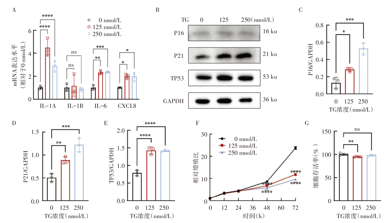
| 基因名称 | 序列 |
|---|---|
| IL-1A | 上游:5'-AATCATCAAGCCTAGGTCAGC-3' |
| 下游:5'-CTTCATCTTGGGCAGTCACA-3' | |
| IL-1B | 上游:5'-GTTGAAAGATGATAAGCCCACT-3' |
| 下游:5'-GTTATATCCTGGCCGCCTT-3' | |
| IL-6 | 上游:5'-CACACAGACAGCCACTCACC-3' |
| 下游:5'-ATTTTCACCAGGCAAGTCTCC-3' | |
| CXCL8 | 上游:5'-CAGTTTTGCCAAGGAGTGCTA-3' |
| 下游:5'-TTTTCCTTGGGGTCCAGACA-3' | |
| GAPDH | 上游:5'-GGAGCGAGATCCCTCCAAAAT-3' |
| 下游:5'-GGCTGTTGTCATACTTCTCATGG-3' |

| [1] |
Johnson JA, Tough S. No-271-Delayed Child-Bearing[J]. J Obstet Gynaecol Can, 2017, 39(11):e500-e515. doi: 10.1016/j.jogc.2017.09.007.
pmid: 29080737 |
| [2] | Broekmans FJ, Knauff EA, te Velde ER, et al. Female reproductive ageing: current knowledge and future trends[J]. Trends Endocrinol Metab, 2007, 18(2):58-65. doi: 10.1016/j.tem.2007.01.004. |
| [3] | Levine ME, Lu AT, Chen BH, et al. Menopause accelerates biological aging[J]. Proc Natl Acad Sci U S A, 2016, 113(33):9327-9332. doi: 10.1073/pnas.1604558113. |
| [4] |
Muka T, Oliver-Williams C, Kunutsor S, et al. Association of Age at Onset of Menopause and Time Since Onset of Menopause With Cardiovascular Outcomes, Intermediate Vascular Traits, and All-Cause Mortality: A Systematic Review and Meta-analysis[J]. JAMA Cardiol, 2016, 1(7):767-776. doi: 10.1001/jamacardio.2016.2415.
pmid: 27627190 |
| [5] | Kordus RJ, LaVoie HA. Granulosa cell biomarkers to predict pregnancy in ART: pieces to solve the puzzle[J]. Reproduction, 2017, 153(2):R69-R83. doi: 10.1530/REP-16-0500. |
| [6] | Liu C, Zuo W, Yan G, et al. Granulosa cell mevalonate pathway abnormalities contribute to oocyte meiotic defects and aneuploidy[J]. Nat Aging, 2023, 3(6):670-687. doi: 10.1038/s43587-023-00419-9. |
| [7] |
Tatone C, Amicarelli F. The aging ovary--the poor granulosa cells[J]. Fertil Steril, 2013, 99(1):12-17. doi: 10.1016/j.fertnstert.2012.11.029.
pmid: 23273984 |
| [8] | Chen X, Kang R, Kroemer G, et al. Ferroptosis in infection, inflammation, and immunity[J]. J Exp Med, 2021, 218(6):e20210518. doi: 10.1084/jem.20210518. |
| [9] |
Meng Y, Sun HY, He Y, et al. BET inhibitors potentiate melanoma ferroptosis and immunotherapy through AKR1C2 inhibition[J]. Mil Med Res, 2023, 10(1):61. doi: 10.1186/s40779-023-00497-1.
pmid: 38049916 |
| [10] | Zhao WP, Wang HW, Liu J, et al. Mitochondrial respiratory chain complex abnormal expressions and fusion disorder are involved in fluoride-induced mitochondrial dysfunction in ovarian granulosa cells[J]. Chemosphere, 2019, 215:619-625. doi: 10.1016/j.chemosphere.2018.10.043. |
| [11] |
Wang F, Liu Y, Ni F, et al. BNC1 deficiency-triggered ferroptosis through the NF2-YAP pathway induces primary ovarian insufficiency[J]. Nat Commun, 2022, 13(1):5871. doi: 10.1038/s41467-022-33323-8.
pmid: 36198708 |
| [12] | Ma J, Chen S, Liu J, et al. Cryptochrome 1 regulates ovarian granulosa cell senescence through NCOA4-mediated ferritinophagy[J]. Free Radic Biol Med, 2024, 217:1-14. doi: 10.1016/j.freeradbiomed.2024.03.015. |
| [13] |
Babayev E, Duncan FE. Age-associated changes in cumulus cells and follicular fluid: the local oocyte microenvironment as a determinant of gamete quality[J]. Biol Reprod, 2022, 106(2):351-365. doi: 10.1093/biolre/ioab241.
pmid: 34982142 |
| [14] | Hennet ML, Combelles CM. The antral follicle: a microenvironment for oocyte differentiation[J]. Int J Dev Biol, 2012, 56(10/11/12):819-831. doi: 10.1387/ijdb.120133cc. |
| [15] |
Wang S, Zheng Y, Li J, et al. Single-Cell Transcriptomic Atlas of Primate Ovarian Aging[J]. Cell, 2020, 180(3):585-600.e19. doi: 10.1016/j.cell.2020.01.009.
pmid: 32004457 |
| [16] |
Dixon SJ, Lemberg KM, Lamprecht MR, et al. Ferroptosis: an iron-dependent form of nonapoptotic cell death[J]. Cell, 2012, 149(5):1060-1072. doi: 10.1016/j.cell.2012.03.042.
pmid: 22632970 |
| [17] | Hu J, Wang H, Fang J, et al. Ovarian aging-associated downregulation of GPX4 expression regulates ovarian follicular development by affecting granulosa cell functions and oocyte quality[J]. FASEB J, 2025, 39(6):e70469. doi: 10.1096/fj.202401580RR. |
| [18] | Chen H, Wang S, Ding Z, et al. Rbbp7-mediated deacetylation of Acsl4 promotes ovarian aging by enhancing ferroptosis[J]. Int J Biol Macromol, 2026, 339(Pt 2):149976. doi: 10.1016/j.ijbiomac.2025.149976. |
| [19] | Chen D, Wang Y, Wang X, et al. Corrigendum to "Nuclear receptor coactivator 4 linked to follicular dysplasia in polycystic ovary syndrome: A key regulator that aggravates ovarian granulosa cells ferritinophagy and ferroptosis" [Biochim Biophys Acta Mol Basis Dis. 1871 (7) (Oct 2025) 167955 (BBADIS 167955)][J]. Biochim Biophys Acta Mol Basis Dis, 2026, 1872(5):168183. doi: 10.1016/j.bbadis.2026.168183. |
| [20] | Hu L, Hong T, He Y, et al. Chromosome Segregation-1-like Gene Participates in Ferroptosis in Human Ovarian Granulosa Cells via Nucleocytoplasmic Transport[J]. Antioxidants(Basel), 2024, 13(8):911. doi: 10.3390/antiox13080911. |
| [21] |
Qin X, Zhao Y, Zhang T, et al. TrkB agonist antibody ameliorates fertility deficits in aged and cyclophosphamide-induced premature ovarian failure model mice[J]. Nat Commun, 2022, 13(1):914. doi: 10.1038/s41467-022-28611-2.
pmid: 35177657 |
| [22] | Zhou C, Guo Q, Lin J, et al. Single-Cell Atlas of Human Ovaries Reveals The Role Of The Pyroptotic Macrophage in Ovarian Aging[J]. Adv Sci(Weinh), 2024, 11(4):e2305175. doi: 10.1002/advs.202305175. |
| [1] | WEI Yuan-jie, YUAN Li-hua, SUN Zhen-gao. Immunological Mechanism of Quality Decline in Elderly Oocytes [J]. Journal of International Reproductive Health/Family Planning, 2026, 45(2): 145-149. |
| [2] | KONG Jing, YU Lan, ZHANG Cui-lian. The Impact of Vitamin D on Assisted Reproductive Technology Outcomes in Women with Polycystic Ovary Syndrome [J]. Journal of International Reproductive Health/Family Planning, 2026, 45(2): 154-159. |
| [3] | WANG Hai-yun, WEI Jia-yu, LAN Tian-ning, ZHANG Ke-xin, ZHANG Hui-ying, TIAN Wen-yan. Research Progress on Amino Acid Metabolism and Polycystic Ovary Syndrome [J]. Journal of International Reproductive Health/Family Planning, 2026, 45(2): 160-165. |
| [4] | LIN Kai-li, LIU Yin, WANG Jiao-jian, SONG Dian-rong, ZHANG Wei, LU Di. Animal Experimental Study on the Efficacy of Traditional Chinese Medicine Syndrome Differentiation Treatment for PCOS Based on Core Indicator Target Evaluation [J]. Journal of International Reproductive Health/Family Planning, 2026, 45(2): 89-96. |
| [5] | LIU Ying, NING Shu-ting, ZHANG Chun-ren, DAI Fang, MO Hui-ying, MA Hong-xia. Effect of Prenatal Intrauterine Exposure to High Anti-Müllerian Hormone and Androgen on the Gut Microbiota of Offspring Mice [J]. Journal of International Reproductive Health/Family Planning, 2026, 45(2): 97-103. |
| [6] | KANG Xu-li, LIU Bo-xin, HE Xiao, ZHAI Hui. Age-Period-Cohort Analysis of the Disease Burden of Polycystic Ovary Syndrome in China from 1990 to 2021 and Prediction of the Trend [J]. Journal of International Reproductive Health/Family Planning, 2026, 45(1): 11-17. |
| [7] | CHEN Wen-xin, YU Chi-yuan, XU Bo-qun. Advances in Clinical and Basic Research on the Transgenerational Inheritance of Polycystic Ovary Syndrome [J]. Journal of International Reproductive Health/Family Planning, 2026, 45(1): 60-66. |
| [8] | LIN Tuo, NING Shu-ting, HUA Ying, YE Li-hua, MA Hong-xia. Research Progress on Anti-Müllerian Hormone and Inhibin B in the Transgenerational Effects of Polycystic Ovary Syndrome [J]. Journal of International Reproductive Health/Family Planning, 2026, 45(1): 66-70. |
| [9] | ZHANG Xi-ruo, WANG Rong-rong, SU Jing, XUE Feng-xia. The Role of Th17 Cells and IL-17A in the Pathogenesis of Premature Ovarian Insufficiency [J]. Journal of International Reproductive Health/Family Planning, 2025, 44(6): 501-505. |
| [10] | LENG Ya-wen, SHI Bai-chao, WANG Yu, WU Xiao-ke. The Relationship between Adipokines and Polycystic Ovary Syndrome [J]. Journal of International Reproductive Health/Family Planning, 2025, 44(6): 518-523. |
| [11] | LI Meng-yuan, GAO Zheng, XU Xin. Mechanism of Yishen Quzhuo Formula in Treating PCOS-IR Based on Network Pharmacology and Animal Experiments [J]. Journal of International Reproductive Health/Family Planning, 2025, 44(5): 353-360. |
| [12] | CHAI Ling-na, SHI Jie, LI Yan-li, GAO Han, CHENG Shi-yu, OUYANG Xi-yan. A Case of Uterine Tumor Resembling Ovarian Sex Cord Tumor with Adenomyosis [J]. Journal of International Reproductive Health/Family Planning, 2025, 44(5): 383-387. |
| [13] | YU Jian-nan, GAO Zhu-wei, GE Hang, LIU Yang, SHI Bai-chao, BAI Yu-xin, GAO Jing-shu, WANG Yu, WU Xiao-ke. The Potential Therapeutic Role of Quercetin in Polycystic Ovary Syndrome [J]. Journal of International Reproductive Health/Family Planning, 2025, 44(5): 393-398. |
| [14] | NIU Jia-jia, WANG Yu, WU Xiao-ke. Research Progress on Probiotic Therapy for Polycystic Ovary Syndrome [J]. Journal of International Reproductive Health/Family Planning, 2025, 44(5): 399-404. |
| [15] | XIONG Min, YANG Dan-dan, CHAI Meng-han, ZHANG Qian-nan, ZHANG Zhi-guo, CHEN Bei-li. Effects of Per- and Polyfluoroalkyl Substances on Female Reproductive Health and Outcomes of Assisted Reproductive Technology [J]. Journal of International Reproductive Health/Family Planning, 2025, 44(5): 411-415. |
| Viewed | ||||||
|
Full text |
|
|||||
|
Abstract |
|
|||||